1. The Spatial Dimension of Genetic Disease: Moving Beyond 1D Variant Calling
For decades, the dominant paradigm in clinical genetics and molecular biology treated chromosomes as one-dimensional linear strings of nucleotides. Variant interpretation pipelines concentrated almost exclusively on protein-coding sequences, prioritizing missense substitutions, nonsense mutations, and frameshift indels, while categorizing noncoding structural variants (SVs) primarily as copy number alterations (CNVs) that dictate linear gene dosage. Yet comprehensive whole-genome sequencing has revealed that more than 95% of disease-associated variants identified through genome-wide association studies (GWAS) reside entirely within noncoding genomic intervals. Inside the eukaryotic nucleus, approximately two meters of genomic DNA are compacted within a space measuring mere micrometers across. This compaction is not random polymer entanglement; rather, it is folded into an exquisite, hierarchical three-dimensional architecture that dictates spatial proximity between distal cis-regulatory elements and their target gene promoters. As established across recent genomic studies, structural variants, including deletions, tandem duplications, inversions, translocations, and complex catastrophic rearrangements such as chromothripsis, exert profound phenotypic consequences by directly disrupting this higher-order architecture. When an SV removes an insulating architectural boundary, inverts a directional motif, or relocates an active super-enhancer across domain borders, the spatial insulation of the nucleus is breached. Promoters are ectopically activated in a process termed enhancer hijacking, while physiological regulatory contacts are severed. Consequently, decoding the pathogenicity of structural variation requires transitioning from linear sequence alignment to 3D topological genomics. Before examining specific molecular case studies, contrasting the classical gene-centric view with the 3D topological framework illustrates how spatial genome architecture redefines our understanding of genomic lesions: Analytical Dimension,Classical Gene-Centric Framework,3D Topological Genomics Framework Primary Genomic Focus,"Exons, splice junctions, and protein-coding reading frames","Architectural boundaries, noncoding regulatory elements, and chromatin loops" Pathogenic Mechanism,"Amino acid substitution, truncated protein, or linear gene dosage alterations","Enhancer hijacking, TAD fusion, neo-TAD creation, and topological position effects" Spatial Coordinate Model,One-dimensional physical distance along the chromosome (base pairs),Contact frequency and three-dimensional Euclidean distance in nuclear space Interpretation of Noncoding SVs,Frequently classified as variants of uncertain significance (VUS),Evaluated for disruption of insulated neighborhoods and ectopic looping Diagnostic Sensitivity,Misses pathogenic rearrangements where coding gene bodies remain intact,Identifies distal enhancer repositioning and structural boundary collapses
2. The Architectural Hierarchy: From Nuclear Territories to Microcompartments and Enhancer Hubs
Spatial genome organization is organized across distinct scales, establishing an interconnected hierarchy that governs transcription, DNA replication timing, and genomic stability. Chromosome Territories and A/B Compartments At the nuclear scale, individual chromosomes occupy discrete, largely mutually exclusive chromosome territories. At megabase resolution, chromosome conformation capture (Hi-C) contact maps partition chromatin into two primary compartmental domains that reflect transcriptional activity and epigenetic state: Compartment A : Transcriptionally active, gene-dense, accessible chromatin characterized by active histone modifications such as H3K27ac, H3K4me1, and H3K4me3. Compartment B : Transcriptionally repressed, heterochromatic, gene-poor regions associated with repressive marks including H3K9me3 (constitutive heterochromatin) and H3K27me3 (facultative heterochromatin), frequently localized near the nuclear lamina or nucleolus. In malignancies, somatic structural alterations and chromatin remodeler mutations frequently induce large-scale compartment switching. A transition from Compartment B to Compartment A can ectopically activate dormant oncogenes, whereas Compartment A-to-B transitions can silence crucial tumor suppressor programs. Topologically Associating Domains (TADs) and Insulated Neighborhoods Subdividing compartments are Topologically Associating Domains (TADs), megabase- to sub-megabase-scale regulatory units characterized by high internal interaction frequencies and strict spatial insulation from adjacent domains. TAD boundaries act as physical barriers, enriched for the zinc-finger insulator protein CTCF and the multiprotein ring complex cohesin. By restricting enhancer-promoter communication largely within domain interiors, TADs ensure that potent regulatory elements activate only their cognate target genes. TAD boundaries are conserved across mammalian species and cell types, reflecting fundamental selective constraints against aberrant inter-domain cross-talk. An integrative catalog generated in human lymphoblastoid cell lines by the Human Genome Structural Variation Consortium identified 18,865 TADs, including 4,596 nested sub-TADs, revealing that 185 structural variants directly alter these chromatin architectures, with 34 intersecting candidate cis-regulatory elements (cCREs). Microcompartments, Stripes, and Multi-Enhancer Hubs Recent technological advances, including Region Capture Micro-C (RCMC) and single-cell Micro-C (scMicro-C), have resolved structural features nested beneath classical TADs: Promoter-Enhancer Stripes (PES) : Continuous directional interaction patterns generated by cohesin complexes that physically reel distal regulatory elements toward an active promoter. Microcompartments and Regulatory Hubs : Fine-scale spatial clusters wherein multiple distal enhancers, Mediator complexes, transcription factors, and RNA Polymerase II coalesce simultaneously around a single promoter. These multi-enhancer hubs provide robust, coordinated transcriptional bursts while preserving stringent regulatory specificity.
3. Biophysics of Chromatin Folding: Loop Extrusion, CTCF Directionality, and Polymer Dynamics
The formation and maintenance of chromatin loops and domain boundaries are governed by active molecular machinery operating in concert with fundamental polymer physics principles. The Loop Extrusion Engine Under the loop extrusion model, the ring-shaped cohesin complex (comprising SMC1, SMC3, RAD21, and STAG proteins) is dynamically loaded onto chromatin fibers by the NIPBL-MAU2 complex. Once engaged, cohesin acts as a molecular motor, extruding bidirectional loops of DNA until it encounters architectural obstacle boundaries. Loop release and turnover are tightly regulated by the release factor WAPL, balancing loop longevity with dynamic chromatin plasticity. Experimental degradation of cohesin eliminates all loop domains across the genome, while depletion of WAPL leads to hyper-extended chromatin loops, demonstrating that domain formation is a dynamic equilibrium between continuous extrusion and programmed release. The Convergent Motif Rule CTCF possesses an asymmetrical, 20-base-pair consensus sequence. Loop extrusion ceases stably when the cohesin ring encounters CTCF molecules bound in a convergent orientation (motifs facing each other). Divergent or tandem parallel motifs fail to halt cohesin extrusion with equal efficiency, permitting loop extension into neighboring territories. Consequently, sequence variations that invert, mutate, or epigenetically hypermethylate a single convergent CTCF binding site can permit cohesin to bypass the boundary, systematically reshaping the surrounding 3D loop landscape. Polymer Physics and the PRISMR Framework Chromatin behaves as a semi-flexible self-avoiding polymer chain subject to thermal fluctuations and molecular crowding. As demonstrated by Nicodemi and colleagues through the PRISMR (Polymer-physics-based Reconstruction of In Situ 3D Chromatin from Contact Maps) framework, genome folding can be modeled using statistical mechanics and minimal energy states. By treating interaction frequencies derived from Hi-C or ChIA-PET as proxies for thermodynamic equilibrium distances, polymer models demonstrate how discrete physical interactions deconvolve complex genomic loci. Crucially, PRISMR demonstrated that structural variations do not merely delete linear sequence; they trigger widespread thermodynamic re-equilibration, producing predictable ectopic contacts and global topological rewiring. In simulations of the EPHA4 locus, PRISMR successfully predicted pathogenic ectopic interactions across both homozygous and heterozygous deletions, aligning with experimental Hi-C contacts generated from mouse limb buds and patient-derived fibroblasts.
4. Mechanisms of Topological Disruption: Boundary Deletions, Neo-TADs, and Enhancer Hijacking
When structural variants intersect the 3D genome, they trigger distinct architectural failure modes that rewire long-range transcriptional control:
A. TAD Boundary Deletion and Domain Fusion
The loss of an architectural boundary merges two previously insulated topological domains into a single fused domain. Enhancers within the upstream domain, previously sequestered from the downstream promoter, now freely interact across the fused interval. Classical developmental phenotypes, such as the limb malformations observed at the EPHA4 locus, demonstrate how boundary deletions allow digit enhancers to misroute activation signals to neighboring genes such as WNT6 or PAX3 .
B. Inversions and CTCF Reorientation
Because loop extrusion depends upon CTCF motif polarity, genomic inversions produce subtle yet catastrophic topological rewiring. Inverting a segment containing a CTCF site flips its directional motif. Cohesin now passes through the original boundary position and halts at alternative downstream sites, creating novel loop architectures and ectopically juxtaposing enhancers with unintended targets.
C. Enhancer Hijacking
Enhancer hijacking occurs when a genomic rearrangement repositions an active, lineage-specific enhancer or super-enhancer in spatial proximity to a proto-oncogene that is normally silent or tightly repressed in that cell type. Rather than generating a chimeric fusion oncoprotein, enhancer hijacking unleashes massive, monoallelic overexpression of the intact wild-type transcription factor or kinase.
D. Neo-TAD Creation and Extrachromosomal Circular DNA (ecDNA)
Complex rearrangements, such as tandem duplications or interchromosomal translocations, can assemble entirely novel chromatin neighborhoods called neo-TADs . In aggressive malignancies, oncogenes and strong enhancers frequently excise from chromosomes into extrachromosomal circular DNA (ecDNA or eccDNA). Free from linear chromosomal constraints, ecDNAs form ultra-dense, interconnected regulatory hubs, amplifying oncogene transcription to pathological extremes and driving intratumoral heterogeneity.
5. Clinical Archetypes in Cancer: Case Studies from AML, T-ALL, and Brain Tumors
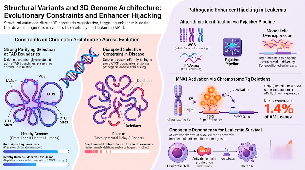
Cancer is increasingly recognized as a disease of both genome sequence and genome architecture. Emerging research highlights how structural rearrangements systematically exploit 3D topology across diverse malignancies: Acute Myeloid Leukemia: Deletion 7q and MNX1 Activation In complex karyotype acute myeloid leukemia (ckAML), a high-risk subtype characterized by multiple copy number alterations, TP53 mutations, and dismal clinical outcomes, deletions of chromosome arm 7q (del(7q)) are among the most common structural lesions, present in nearly 70% of cases. Classically, del(7q) was assumed to act solely via haploinsufficiency of putative tumor suppressors such as CUX1 or MLL3 . However, systematic profiling of 39 ckAML clinical trial samples with the computational discovery algorithm pyjacker revealed a completely different pathomechanism: In samples harboring del(7)(q22q36), the interstitial deletion excises intervening chromosomal DNA, juxtaposing a potent hematopoietic super-enhancer from the CDK6 region directly adjacent to motor neuron and pancreas homeobox 1 ( MNX1 ), located at 7q36.3 outside the common deleted region. MNX1 is normally completely repressed in adult hematopoietic stem and progenitor cells. This deletion-induced enhancer hijacking drives severe monoallelic MNX1 overexpression in approximately 1.4% of AML patients, co-occurring significantly with BCOR mutations. Functional validation using circular chromosome conformation capture (4C-seq) confirmed direct physical interaction between MNX1 and the CDK6 locus across primary AML and patient-derived xenograft (PDX) samples. To functionally validate this mechanism, researchers inserted a 1-kb candidate enhancer from the CDK6 locus upstream of MNX1 in ChiPSC22 induced pluripotent stem cells using CRISPR/Cas9 homology-directed repair; upon hematopoietic differentiation, the engineered cells exhibited significant MNX1 activation. Furthermore, shRNA-mediated knockdown of MNX1 in PDX competition assays severely impaired leukemic cell fitness, demonstrating that hijacked MNX1 expression is strictly essential for leukemia propagation. Beyond MNX1 , pyjacker uncovered MECOM activation driven by GATA2 enhancer hijacking via chromothripsis or complex translocations on chromosome 3, as well as aberrant activation of the erythropoietin gene ( EPO ) via an extrachromosomal circular DNA fusing chromosomes 7 and 11 in acute erythroleukemia. T-Cell Acute Lymphoblastic Leukemia: TAL1 Neighborhood Disruption In T-ALL, microdeletions upstream of the TAL1 proto-oncogene remove an insulated CTCF boundary that segregates the TAL1 promoter from a super-enhancer located near the CMPK1 promoter. Computational modeling with 3D-GNOME and physical 3D distance calculations show that boundary deletion collapses the insulated neighborhood, pulling the TAL1 promoter and the CMPK1 enhancer into close Euclidean proximity and driving oncogenic T-cell transformation. Medulloblastoma and Glioma: GFI1 and PDGFRA In pediatric medulloblastoma, structural variations near domain borders reposition active enhancers adjacent to GFI1 , GFI1B , or PRDM6 , generating oncogenic regulatory contacts without altering coding exons. In IDH-mutant gliomas, DNA hypermethylation at CTCF binding sites disrupts insulator protein binding, allowing a constitutive enhancer to aberrantly contact and activate the receptor tyrosine kinase oncogene PDGFRA .
6. Evolutionary Constraint and Population Architecture: Purifying Selection at Domain Borders
Is 3D chromatin architecture an incidental byproduct of nuclear packaging, or is it subjected to active evolutionary selection? Comprehensive analyses comparing structural variation across evolutionary timescales have yielded profound insights into genome conservation: Signatures of Purifying Selection Across Primates and Healthy Humans By mapping structural variants across great ape divergence (chimpanzees, gorillas, orangutans), the human population (1000 Genomes Project), and disease cohorts, Fudenberg and Pollard quantified the selective pressure acting on 3D chromatin features: In healthy humans and throughout primate evolution, deletions are strongly and systematically depleted at active chromatin states, strong CTCF clusters, and TAD boundaries . The strength of purifying selection scales directly with feature importance: deletions present in multiple healthy individuals are approximately 1.7-fold more depleted at boundaries than unique singletons; boundaries shared across multiple cell types are roughly 1.4-fold more depleted; and boundaries in the top quintile of single-nucleotide evolutionary conservation (phyloP scores) are 2.2-fold more depleted. TAD boundaries are avoided by deletions as strongly as the transcription start sites of intermediately expressed protein-coding genes. This establishes that noncoding structural insulators are subject to purifying selection comparable to essential coding elements. The overall distribution of deletions and duplications in healthy humans rapidly plummets beyond approximately 2 Mb. This 2-Mb threshold coincides with the maximum distance over which mammalian enhancers typically operate, the scale of the largest TADs, and the physical limit over which cohesin enriches contact frequency, demonstrating that the genome is constrained to prevent ectopic inter-domain contact. The Breakdown of Selection in Human Disease This signature of evolutionary constraint completely dissolves in pathological cohorts: In patients with autism spectrum disorder and severe developmental delay, deletions occur remarkably uniformly across genomic features , displaying no avoidance of strong CTCF clusters or TAD boundaries. These unconstrained de novo deletions systematically breach insulated neighborhoods, underscoring enhancer hijacking as a widespread cause of neurodevelopmental disorders. In cancer genomes, somatic deletions actually display a slight enrichment at active chromatin features, CTCF clusters, and domain borders, reflecting positive selection for rearrangements that rewire regulatory networks and activate oncogenes. Population-Scale Topological Variability Analyses leveraging 1000 Genomes Project data and lymphoblastoid cell line ChIA-PET maps (incorporating over 44,000 CTCF-cohesin interactions) demonstrate that while the core CTCF scaffold is conserved, individual human genomes harbor thousands of SVs altering local loop topology: Out of 68,818 unique SVs in the 1000 Genomes catalog, 4,944 unique patterns alter CTCF interaction anchors within Chromatin Contact Domains (CCDs). While deletions dominate structural variations in interaction anchors, domain boundaries are surprisingly enriched for duplications . This indicates that boundary duplication may serve as an evolutionary engine for regulatory innovation, allowing one duplicated domain to preserve essential transcriptional control while the second evolves novel spatial promoter-enhancer connections. Mapping expression quantitative trait loci (eQTLs) to 3D contacts reveals that noncoding structural variants influence gene transcription primarily by rearranging local RNA Polymerase II-mediated interaction networks within conserved CTCF-insulated domains.
7. Computational Tools and Algorithms for 3D Genomic Discovery: From Hi-C to PRISMR and pyjacker
Deciphering how structural variants perturb chromatin conformation requires specialized computational workflows that combine high-throughput sequencing with physical and statistical modeling. The primary bioinformatics and biophysical tools designed to uncover and predict the topological consequences of structural variation include: Computational Tool,Primary Data Inputs,Core Algorithm / Framework,Major Application in 3D Genomics pyjacker,WGS + bulk RNA-seq + H3K27ac/P300 ChIP-seq,"Outlier expression modeling, heterozygous SNP monoallelic ratio, and distance-to-breakpoint scoring",Identification of sample-specific and recurrent enhancer hijacking events in cancer cohorts without matched normals PRISMR,Hi-C / 5C contact matrices + structural variant coordinates,Statistical polymer physics and Monte Carlo simulated annealing of energy states,"In silico prediction of chromatin refolding, ectopic loop generation, and TAD reconfiguration induced by SVs" 3D-GNOME 2.0,ChIA-PET (CTCF / RNAPII) + 1000 Genomes SV calls,Graph-based multiscale modeling; PET clusters and singletons optimization,Generation of 3D Euclidean spatial coordinates and visualization of structural variant-altered looping structures NeoLoopFinder,High-resolution Hi-C / Micro-C contact maps,Algorithmic detection of neo-loops across reconstructed rearranged chromosomes,Direct identification of chromatin loops connecting hijacked enhancers and oncogenes in cancer genomes cis-X,WGS + RNA-seq + matched normal samples,Allelic imbalance and regulatory variant mapping across phased haplotypes,Discovery of noncoding driver mutations and enhancer hijacking in individual cancer genomes The Mechanics of pyjacker Traditional eQTL approaches or linear regression tools fail to detect enhancer hijacking events that occur in only one or two patients within a heterogeneous cohort. The pyjacker framework circumvents this limitation through an innovative multi-step scoring pipeline: Candidate Segregation : Samples are partitioned into candidate samples (harboring a breakpoint within the same TAD or within a defined genomic window) and reference samples (lacking local breakpoints). Outlier Expression Scoring : Expression levels in candidate samples are statistically tested against the reference distribution to identify significant overexpression. Monoallelic Expression Verification : In candidate samples with heterozygous SNPs covered by RNA-seq reads, pyjacker calculates the variant allele frequency to ensure overexpression originates exclusively from the rearranged allele. Enhancer Proximity Scoring : Intersecting breakpoint coordinates with cell-type-specific active chromatin marks (H3K27ac, P300) evaluates the regulatory strength of enhancers brought into spatial proximity with the candidate gene.
8. Future Horizons: 4D Nucleome Modeling and Chromatin-Directed Therapeutics
The convergence of chromosome conformation capture, high-resolution imaging, and computational structural genomics is rapidly transforming 3D genome analysis from a descriptive academic discipline into a cornerstone of precision medicine. Emerging Technologies Single-Cell Multimodal Profiling : Techniques such as single-cell Micro-C (scMicro-C) combined with single-cell RNA sequencing now resolve cell-to-cell variability in chromatin folding, distinguishing stable architectural frameworks from transient transcriptional bursts. Micro-Capture-C (MCC) and RCMC : Hybridization-capture methodologies achieve base-pair resolution, mapping physical contacts between transcription factor binding motifs and individual nucleosomes. Deep Learning and Generative 3D Modeling : Neural network architectures trained on high-resolution contact maps and sequence motifs are increasingly capable of predicting in silico how patient-specific structural variants reshape nuclear architecture before experimental validation. Therapeutic Implications Recognizing cancer as a disease of topological dysregulation unlocks novel therapeutic paradigms: Disrupting Oncogenic Super-Enhancers : Small-molecule inhibitors targeting bromodomain proteins (e.g., BRD4) or Mediator subunits destabilize the protein-protein condensates that sustain hijacked enhancer-promoter hubs. Targeting Architectural Machinery : Modulating cohesin loading factors or regulating chromatin accessibility can selectively attenuate aberrant long-range looping in tumor cells. Epigenome Editing : Engineered dCas9-effector complexes can deposit repressive histone methylation (H3K9me3) onto hijacked enhancers or reactivate methylated CTCF boundaries, restoring native topological insulation. By integrating physical modeling, population genetics, and machine learning, computational genome biology is unraveling the rules of spatial nuclear architecture, paving the way for precision diagnostics that interpret genetic disease through its true three-dimensional nature.